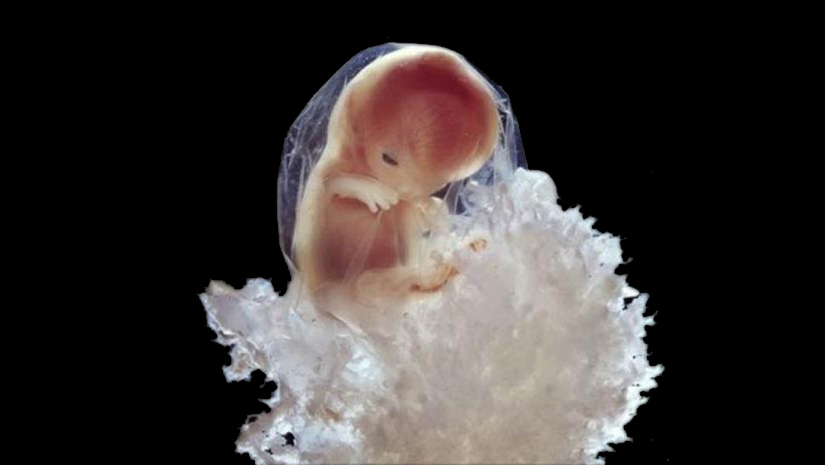
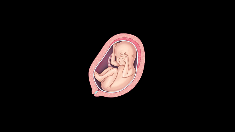
Swedish photographer and scientist Lennart Nilsson in 1965, with his conventional cameras took enthralling and incredible photographs taken with cameras with macro lenses, an endoscope and scanning electron microscope, Nilsson captured the different stages of a baby being formed in the mother’s womb. The pictures were later collected and published in a hardcover book called A Child Is Born by Jonathan Cape.
Day 1: Sperm in Fallopian Tube

Days 5-6: Blastocyst containing many more cells now enters the womb

Day 8: Embryo attaches to the wall of the womb

5 Weeks: 9mm Embryo. Face beginning to form with openings for eyes, nose and mouth.

5-6 Weeks Embryonic Cells form placenta

8 Weeks: Embryo protected in Foetal sac

10 Weeks: Embryo’s eyelids are semi shut

16 Weeks: Foetus uses its hands to explore body and surroundings

16-18 Weeks: Network of Blood vessels are visible through the skin

18 Weeks: Foetus is 14 cms and can perceive sounds

19 Weeks: Nails start to appear

20 Weeks: Head covered with minute hairs

24 Weeks: Foetus is 3 months old

26 Weeks: Foetus is almost fully grown

28-30 weeks: Baby turns up-side down in the womb

36 Weeks: Baby is ready to take birth

By: Archa Dave


